Diagnostics of Amebic Ocular and Meningoencephalitis Infections
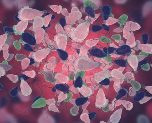
Rare infections of the eye and brain caused by soil and freshwater amebic parasites, namely, Acanthamoeba spp., Balamuthia mandrillaris, and Naegleria fowleri require rapid diagnosis to support targeted treatments and improved patient outcomes. BioGX has recently launched the amebic parasite multiplex for the BD MAX™ system that targets:
- Acanthamoeba spp. (inclusive of genotypes T1-T23)
- Balamuthia mandrillaris
- Naegleria fowleri
A brief overview of clinical manifestations and testing approaches are defined below to highlight the importance of rapid real-time PCR diagnostics of suspected cases of keratitis, Granulomatous Amebic Encephalitis (GAE) and Primary Amebic Meningoencephalitis (PAM). Speed of diagnosis allows for effective use of anti-amoebic drugs, and support of surveillance activities.
Diagnostic Strategies
KeratitisAcanthamoeba spp. keratitis cases are associated with contact lens users. A majority of cases have occurred in North and South America, China, India, Australia and limited cases reported in Europe and Africa. Clinical manifestations include early “ring” infiltrate, pain and perineuritis.To prevent poor prognosis and irreversible blindness¹, rapid testing prevents surgery and improves patient outcomes2,3.
Current diagnostic tests for Acanthamoeba spp. Keratitis:
- PCR of corneal scrapings or biopsy
- In vivo confocal microscopy
- Cultivation
- Histopathology
Amebic MeningoencephalitisAcanthamoeba spp. infections can lead to progressive infection of the CNS (i.e., GAE) that occurs among immunocompromised/immunocompetent individuals, organ transplant recipients, and those with other comorbidities. The typical sites of infection include nasopharyngeal or cutaneous epithelium. Cases of Acanthamoeba GAE have been reported in Australia, Europe, Africa, South America, and the United States4.Clinical manifestations include confusion or other cognitive changes, fever, seizures, difficulty walking, impaired speech, vision or hearing, headache, and nausea/vomiting5,6.
Current diagnostic tests for Acanthamoeba spp. (GAE) :
- Brain scans (CT/MRI)
- PCR of tissue or CSF samples
- Cultivation
- Histopathology
Balamuthia mandrillarisBalamuthia mandrillaris infections can lead to progressive infection of the CNS (i.e., GAE) that occurs among immunocompromised/immunocompetent individuals, organ transplant recipients, and those with other comorbidities. The predominant site of infection is cutaneous epithelium. A majority of cases occur in the warmer climates of Latin America, the southwestern United States, Asia, and Australia, with limited cases reported in Europe. Clinical manifestations include confusion or other changes, fever, seizures, difficulty walking, impaired speech, vision or hearing, headache, and nausea/vomiting⁷.
Current diagnostic tests for Balamuthia mandrillaris (GAE):
- Brain scans (CT/MRI)
- PCR of tissue, skin, or CSF samples
- Immunodiagnostic assays
Naegleria fowleriNaegleria fowleri is the causative agent of the invasive and fulminant, often fatal, form of meningoencephalitis referred to as Primary Amebic Meningoencephalitis (PAM). The site of infection is the olfactory neuroepithelium. Although most cases have been reported in the United States and Pakistan, infections have also occurred across Asia, Mexico, Africa, Europe, and Australasia. Clinical manifestations include headache, fever, nausea, nuchal rigidity, personality changes, seizures, coma, and behavioral abnormalities8,9.
Current diagnostic tests for Naegleria fowleri (PAM):
- Brain scans (CT/MRI)
- PCR of CSF samples
- Histopathology
References
- Zhang Y., Xu X., et al. 2003. The global epidemiology and clinical diagnosis of Acanthamoeba keratitis. Journal of Infection and Public Health 16(6): 841-852.
- Li G. & Shekhawat N. 2022. Acanthamoeba epitheliopathy: Importance of early diagnosis. American Journal of Ophthalmology Case Reports 25(26): 101499.
- Shareef O., Shareef S., et al. 2023. New frontiers in Acanthamoeba keratitis diagnosis and management. Biology 12(12): 1489.
- Marciano-Cabral F., and Cabral, G. 2003. Acanthamoeba spp. as Agents of Disease in Humans. Clinical Microbiology Reviews 16(2): 273–307.
- Kalra S.K., Sharma P., et al. 2020. Acanthamoeba and its pathogenic role in granulomatous amebic encephalitis. Experimental Parasitology 208: 107788.
- Haston J.C., O’Laughlin K., et al. 2023. The epidemiology and clinical features of non-keratitis Acanthamoeba infections in the United States, 1956–2020. Open Forum Infectious Diseases, US: Oxford University Press 10(1): ofac682.
- Bhosale N.K. and Parija S.C. 2021. Balamuthia mandrillaris: An opportunistic, free-living ameba – An updated review. Tropical Parasitology Oct 20; 11(2): 78–88.
- Zhang H., Cheng X. 2021. Various brain-eating amoebae: the protozoa, the pathogenesis, and the disease. Frontiers of Medicine 15(6): 842–866.
- Marri A.R., Hamer D.H., et al. 2025. Naegleria fowleri and the future of surveillance: A one-health call to action. One Health 101215.
Disclaimer: Information in the blog is provided to educate and propagate general awareness and not intended to make any recommendations for diagnosis or treatment of a disease. The reader is encouraged to independently verify the accuracy of information presented in the blog.


